Knees - Pain, Sprains & Meniscus Injuries

The knee is the largest joint in the body. It is subject to a wide variety of traumatic, mechanical and inflammatory disorders. The leading injuries to the knee in both adults and children alike are primarily ligament strains and tears, including the patellar ligament as well as supporting ligaments around the joint. Meniscus injuries are also a common cause of knee pain, accounting for one sixth of knee surgeries. Meniscal damage can be caused by either trauma or gradual degeneration. Tears are the most common form of meniscal injuries, and have poor healing ability primarily because less than 25% of the menisci receive a direct blood supply. Another interesting fact is that the medial meniscus firmly adheres to the deep surface of the medial collateral ligament (MCL), an important stabilizing ligament. Therefore injury to the medial meniscus will very often also result in injury and sprain to the MCL. The cause of the knee pain may be the MCL sprain, but MCL sprains are usually not addressed, especially if the MRI shows a meniscal tear. This could explain pain persisting after meniscal surgery.

MRI’s can be misleading in diagnosing knee pain because they may show abnormalities not related to the patient’s current pain complaint. Many studies have documented the fact that abnormal MRI findings exist in large groups of pain-free individuals. The finding of asymptomatic changes in knee joints during surgery is also not uncommon. One study looked at the value of MRI’s in the treatment of knee injuries and concluded “Overall, magnetic resonance imaging diagnoses added little guidance to patient management and at times provided spurious [false] information.” (LaPrade RF et al American Journal of Sports Medicine. 22(6):739-745) Therefore MRI findings should always be correlated to a patient’s current pain complaint, history and examination.

Conditions which have been successfully treated with Prolotherapy, PRP Prolotherapy and Biocellular (Stem/Stromal Cell) Prolotherapy include: Degenerative Arthritis, Meniscal Tears, Medial Collateral Ligament Sprains, Coronary Ligament Sprains, Pes Anserinus Tendinitis, Patellar Tendonopathy, Patellofemoral Pain Syndrome (PFPS), Lateral Collateral Ligament Sprains and partial ACL tears.


Meniscal Tears

As you can see from this drawing, there is a “medial” (towards the middle of the body) meniscus and a “lateral” (towards the outside of the body) meniscus. The presence of healthy menisci is important for proper functioning of the knee.

Traditional treatment has focused primarily on surgical repair or removal of all or part of the meniscus. Unfortunately, surgery may not always be successful, for a couple of reasons. First, meniscal tissue is very limited in its ability to heal because most of it (the inner two-thirds, which is also where most tears occur[1]) has zero blood supply.[2] Secondly, a meniscal tear may not be the cause of a person’s pain. Most people do not realize that the inner two-thirds of the meniscus, in addition to having no blood supply, has zero nerve fibers, so a meniscus tear there does not actually cause pain. This makes perfect sense when a high number of abnormal MRI findings, such as meniscal tears, exist in people without any pain[3]; in fact, meniscal tears even exist on MRI’s of pain free high functioning professional athletes.[4] So the presence of a meniscal tear on MRI may not necessarily explain what is causing a person’s pain, and so surgery on that tear may not resolve the problem.

What is more likely causing pain in these situations? To answer this, I will explain a little more about how the meniscus helps to provide stability in the knee. A common place for knee pain in on the inner side of the knee, called the medial knee. This is where the medial meniscus connects to the medial collateral ligament (abbreviated “MCL”). In a healthy knee, the medial meniscus and the MCL are firmly attached to each other in order to provide knee stability.

n x-ray image of a runner montaged with images showing the ligament antatomy of the knee.
Meniscal tears are frequent injuries seen by orthopedic medicine physicians. The meniscus is a C-shaped piece of cartilage that provides cushioning in the knee. The word “meniscus” comes from Latin for “crescent moon”, which is what the meniscus looks like. The plural of meniscus is “menisci”.

An illustration of Medial meniscus/medial collateral ligament (MCL) interface, normal anatomy
Medial meniscus/medial collateral ligament (MCL) interface, normal anatomy. As can be seen, the medial collateral ligament is firmly attached to the medial meniscus in order to provide stability in a healthy knee. Disruption of this interface connection can result in instability and pain.

Because these two tissues are connected, whenever there is an injury to any part of the medial meniscus, there will almost always be corresponding microtears and injury to the MCL. This causes a disruption at the interface, eventually resulting in ligament laxity, instability and pain. An MRI is done, which will show the medial meniscus tear, but not usually the corresponding ligament laxity and microtears. Therefore, meniscus surgery can fail because the ligament injury, which may actually be causing the pain, is never addressed. And remember, as discussed previously, that even with minimal surgery, there is an increased risk for the early onset or acceleration of osteoarthritis (OA).[5] Therefore, unless there is a clear-cut surgical need, an alternative nonsurgical approach is desirable.

All three Prolotherapy formulas have been used successfully to stimulate the repair of meniscal tears, in addition to being able to directly treat ligament injury and laxity. Dextrose and Platelet-rich plasma injections have been shown to be effective in many studies,[6] improving pain as well as halting the progression of meniscal damage, as documented by MRI.[7] Biocellular (Stem Cell–Rich) treatment also has the therapeutic potential to directly and indirectly contribute to meniscal healing.[8] In a case report, a patient with an MRI-documented meniscal tear and a two-year history of knee pain received one adipose-derived Biocellular treatment. Three months later, a repeat MRI showed almost complete disappearance of the meniscal tear, along with significant pain reduction.[9] Other research papers agree that the Biocellular therapy has much potential in meniscal treatment, especially the adipose-derived sources.[10]

The science on prolotherapy treatments for the knee


Dextrose

Chondrogenic effect of intra-articular hypertonic-Dextrose (Prolotherapy) in severe knee osteoarthritis (2016)
Improvement in function and pain, as well as increased cartilage, were seen after dextrose prolotherapy, suggesting disease-modifying effects.


Platelet-Rich Plasma

Treatment of degenerative meniscal tear with intrameniscal injection of platelet-rich plasma (PRP) (2020)
Direct injection of PRP into meniscal tears results in improvement in pain/function and MRI imaging.


Biocellular (Stem Cell-Rich) Prolotherapy

Autologous adipose-derived biocellular (Stem Cell-Rich) prolotherapy into Hoffa’s Fat Pad Improves knee osteoarthritis (2021)
A case series demonstrates improvement in quality and density of the fat pad, knee pain reduction, and increased function in patients with moderate-to-severe knee osteoarthritis.


For additional scientific papers and resources on prolotherapy for knee conditions visit the Learning Hub

Case Report: Lifetime Bodybuilder

Barry Taft lifting weights

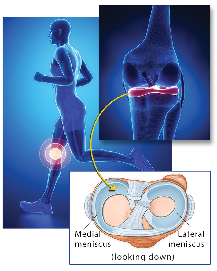
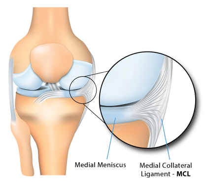
At age 26, I underwent ACL (anterior cruciate ligament) reconstruction knee surgery for an injury sustained during a gymnastics training session. After two years of rehab, I was able to return to gymnastics competition for a couple more years. I then took a 14-year hiatus, returning to weightlifting and fitness training at age 44. When I started training again, it became clear that there was something wrong with my knee. I was told I suffered from patellar Tendinitis due in part to the surgical technique used in my ACL operation years earlier, which utilized the central portion of my patellar tendon as the replacement for the ACL (modified Jones technique). This Tendinitis became worse as I continued to train, and I developed compensatory additional joint pain and symptoms, primarily in the iliotibial band and its attachments around my knee.

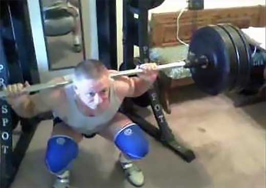
One day, I was squatting 400 pounds and my knee wobbled. There was no immediate pain, and I was able to rack the bar, but two weeks later, while on the leg press, after just two repetitions with a moderate weight, I felt a sharp pain on the outside of the right knee and heard a loud snap. From that point on, my right knee continued to suffer, and my leg started to get progressively weaker. After a year of trying rest and rehab, it was no better.

I had heard of Prolotherapy and finally decided to give it a try; after all, I had very little to lose. At that point, I was not able to do the sport I loved and had tried everything else. Since seeing Dr. Alderman and receiving Prolotherapy, my knee has gotten progressively better. She began conservatively with a few Dextrose Prolotherapy treatments, followed by three PRP Prolotherapy treatments, and then one Biocellular (Stem Cell–Rich) Prolotherapy treatment using my fat as the source of cells. So in case anyone asks, yes, I have had liposuction!

My knee has made steady progress and continues to be good, pain free, and stable over three years since my last treatment. I just recently squatted 400 pounds with no problems. That was my touchstone to know if my knee had been rehabilitated. I would say to anyone who asks that it has! I am very grateful.

Barry Taft
Lifetime Bodybuilder

Tali is a dog that had her knee successfully treated with PRP and Biocellular (Stem Cell-Rich) Prolotherapy.
This short video also addresses a commonly asked question, "Is PRP a placebo?""

The evidence speaks for itself! It is well agreed in medicine that animals do not experience this placebo phenomena because they have no expectations of improvement.

Bibliography

[1] Kimura M, Shirakura K, Hasegawa A et al. Second look arthroscopy after meniscal repair. Factors affecting the healing rate. Clinical Orthopedics and Related Research 1995; 314: 185–191.


[2] Cengiz IF, Silva-Correia J, Pereira H, Espregueira-Mendes J, Oliveira JM, Reis RL. Basics of the Meniscus. In Regenerative Strategies for the Treatment of Knee Joint Disabilities (pp. 237–247). Switzerland: Springer International Publishing. 2017.


[3] Bhattacharyya T, Gale D, Dewire P, Totterman S, Gale ME, McLaughlin S, Einhorn TA, Felson DT. The clinical importance of meniscal tears demonstrated by magnetic resonance imaging in osteoarthritis of the knee. The Journal of Bone & Joint Surgery. 2003 Jan 1; 85(1): 4–9; Kornick J, Trefelner E, McCarthy S, Lange R, Lynch K, Jokl P. Meniscal abnormalities in the asymptomatic population at MR imaging. Radiology. 1990 Nov; 177(2): 463–465; Beattie KA, Boulos P, Pui M, O’Neill J, Inglis D, Webber CE, Adachi JD. Abnormalities identified in the knees of asymptomatic volunteers using peripheral magnetic resonance imaging. Osteoarthritis and Cartilage. 2005 Mar 31; 13(3): 181–186.


[4] Kaplan LD, Schurhoff MR, Selesnick H, Thorpe M, Uribe JW. Magnetic resonance imaging of the knee in asymptomatic professional basketball players. Arthroscopy: The Journal of Arthroscopic & Related Surgery. 2005 May 31; 21(5): 557–561.


[5] Chahla J, Gannon J, Moatshe G, LaPrade RF. Outside-in Meniscal Repair: Technique and Outcomes. In The Menisci (pp. 129–135). Heidelberg, Germany: Springer. 2017; Rongen JJ, Rovers MM, van Tienen TG, Buma P, Hannink G. Increased risk for knee replacement surgery after arthroscopic surgery for degenerative meniscal tears: a multi-center longitudinal observational study using data from the osteoarthritis initiative. Osteoarthritis and Cartilage. 2017 Jan 31; 25(1): 23–29.

[6] Hauser RA, Phillips HJ, Maddela HS. The Case for Utilizing Prolotherapy as First-Line Treatment for Meniscal Pathology. Journal of Prolotherapy. 2010; 2(3): 416–437; Braun HJ, Wasterlain AS, Dragoo JL. The use of PRP in ligament and meniscal healing. Sports Medicine and Arthroscopy Review. 2013 Dec 1; 21(4): 206–201; Hauser RA, Phillips HJ, Maddela H. Platelet rich plasma Prolotherapy as first-line treatment for meniscal pathology. Practical Pain Management. 2010 Jul; 10(6): 53–64


[7] Blanke F, Vavken P, Haenle M, von Wehren L, Pagenstert G, Majewski M. Percutaneous injections of platelet rich plasma for treatment of intrasubstance meniscal lesions. Muscles, Ligaments and Tendons Journal. 2015 Jul; 5(3): 162–166.


[8] Yu H, Adesida AB, Jomha NM. Meniscus repair using mesenchymal stem cells–a comprehensive review. Stem Cell Research & Therapy. 2015 Apr 30; 6(1): 86.


[9] Pak J, Lee JH, Lee SH. Regenerative repair of damaged meniscus with autologous adipose tissue-derived stem cells. BioMed Research International. 2014 Jan 30; 2014: Article ID 436029, 10 pages.


[10] Pak J, Lee JH, Park KS, Jeon JH, Lee SH. Potential use of mesenchymal stem cells in human meniscal repair: current insights. Open Access Journal of Sports Medicine. 2017; 8: 33; Striano RD, Battista V, Bilboo N. Non-responding knee pain with osteoarthritis, meniscus and ligament tears treated with ultrasound guided autologous, micro-fragmented and minimally manipulated adipose tissue. Open Journal of Regenerative Medicine. 2017 Jun 27; 6(02): 17; Whitehouse MR, Howells NR, Parry MC, Austin E, Kafienah W. Brady K, Goodship AE, Eldridge JD, Blom AW and Hollander AP, 2017. Repair of torn avascular meniscal cartilage using undifferentiated autologous mesenchymal stem cells: From in vitro optimization to a first‐in‐human study. Stem Cells Translational Medicine; 6(4): 1237–1248.